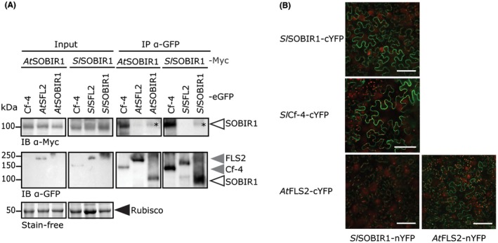
Figure 2.

SOBIR1 constitutively forms homodimers in planta. (A) Myc‐tagged versions of AtSOBIR1 and SlSOBIR1 co‐immunoprecipitate with eGFP‐tagged versions of AtSOBIR1 and SlSOBIR1 (asterisks), respectively, and with Cf‐4‐eGFP, but not with Flagellin‐Sensing 2 (FLS2)‐eGFP. Co‐agroinfiltrations of the various affinity‐tagged proteins were performed in combination with P19 in N. benthamiana leaves at an optical density at OD600 of 0.6 for each construct. Leaves were harvested at 2 dpi, and subjected to IP using anti‐GFP beads, followed by immune blotting (IB). The Rubisco band of the input shows equal loading. The experiment was performed twice and representative results are shown. It should be noted that, because of the low accumulation levels, not all proteins are visible in the input samples. (B) A split‐YFP (yellow fluorescent protein) experiment shows the interaction between SlSOBIR1‐nYFP and SlSOBIR1‐cYFP at the plasma membrane. Leaves of N. benthamiana were agroinfiltrated with constructs driving expression of the indicated constructs, and analysed for interaction at 2 dpi using confocal microscopy. Chloroplast autofluorescence is depicted in red. White bars represent 100 µm. The experiment was performed three times and representative photographs are shown. [Colour figure can be viewed at wileyonlinelibrary.com]
