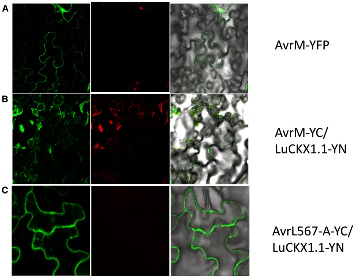
Figure 3.

Subcellular localization of AvrM and bimolecular fluorescence complementation (BiFC) assays with LuCKX1.1‐YN and AvrL567‐YC or AvrM‐YC. Confocal images of flax leaves transiently expressing AvrM‐YFP (A), AvrM‐YC and LuCKX1.1‐YN (B), and AvrL567‐A‐YC and LuCKX1.1‐YN (C). Images in (A) were taken at 4 days post‐inoculation (dpi) and in (B) and (C) at 3 dpi. Left panels, YFP signal; middle panels, chloroplast fluorescence channel; right panels, overlay of YFP, chloroplast and bright field images. YFP, yellow fluorescent protein. [Colour figure can be viewed at wileyonlinelibrary.com]
