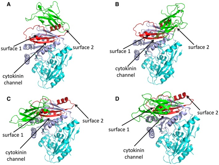
Figure 6.

LuCKX1.1–AvrL567‐A docking studies. The structural models in (A), (B), (C) and (D) represent the top four docking models detected by GRAMM‐X for the proteins LuCKX1.1 and AvrL567‐A (green). The flavin adenine dinucleotide (FAD)‐binding and substrate‐binding domains of LuCKX1.1 are shown in cyan and light blue, respectively. The identified minimal binding region in LuCKX1.1 is coloured red. Black arrows indicate surface 1, surface 2 and the cytokinin channel. [Colour figure can be viewed at wileyonlinelibrary.com]
