Figure 2.

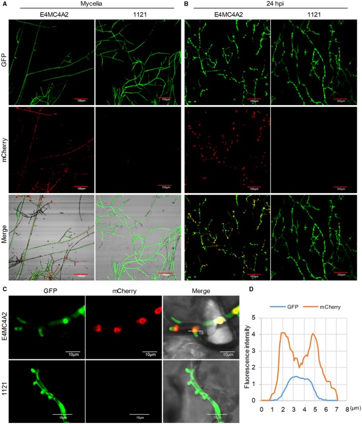
PpE4 accumulates around haustoria after secretion during Phytophthora parasitica infection. (A) Confocal images of mycelia cultured on 5% carrot juice agar medium. The red fluorescence was distributed throughout the mycelial cytoplasm of E4MC4A2 [a transformant expressing cytoplasmic green fluorescent protein (GFP) and full‐length PpE4 (E4FL)‐mCherry], but was not detected in strain 1121 (stably expressing cytoplasmic GFP). (B) Nicotiana benthamiana leaves infected with E4MC4A2 and 1121 were observed by confocal microscopy at 24 h post‐inoculation (hpi). A strong red fluorescence signal was highly accumulated in haustoria, but not in hyphae, during E4MC4A2 infection, whereas GFP fluorescence was evenly distributed in hyphae. No red fluorescence was observed in strain 1121. (C) A magnified lateral view of haustoria showing red fluorescence focused on the outside of the haustoria base and the GFP signal distributed throughout hyphae and haustoria. (D) The fluorescence intensities of GFP and mCherry across the haustorium indicated by the white line labelled ‘2’ in (C). Identical images were obtained from more than 10 haustoria in three independent biological replicates. [Colour figure can be viewed at wileyonlinelibrary.com]
