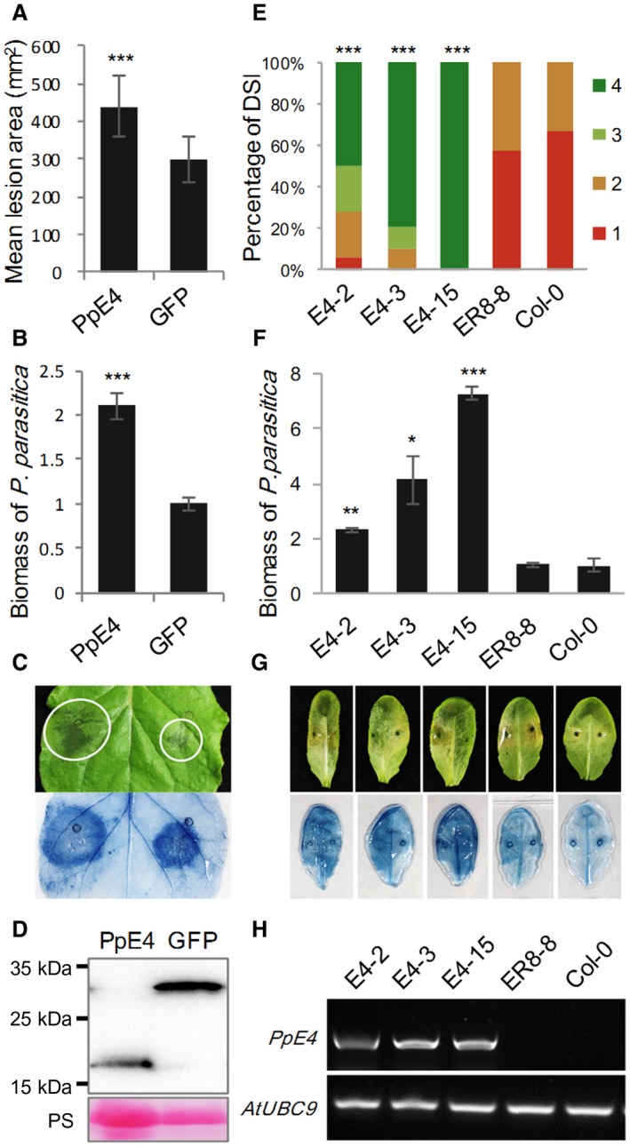
Figure 5.

Heterologous expression of PpE4 renders Nicotiana benthamiana and Arabidopsis more susceptible to Phytophthora parasitica infection. (A) Mean lesion areas were measured at 48 h post‐inoculation (hpi). Agrobacterium tumefaciens strains carrying PpE4 or GFP [optical density at 600 nm (OD600) = 0.01] were infiltrated into different sides of the same leaf, 1 day before inoculation of strain 1121. Error bars represent the standard deviation (SD) of 15 leaves, and asterisks denote significant differences from the green fluorescent protein (GFP) control (two‐tailed t‐test: ***P < 0.001). (B) Quantification of P. parasitica biomass in infected N. benthamiana leaves. Bars represent PpUBC levels relative to NbF‐box levels with SD of three biological replicates. Asterisks denote significant differences from the GFP control (two‐tailed t‐test: ***P < 0.001). (C) A typical leaf photographed and stained by trypan blue. White circles outline the water‐soaked lesions. (D) Protein accumulation was determined at 3 days post‐infiltration (dpi) by western blot using anti‐Flag antibody. Protein loading is indicated by Ponceau stain (PS). Similar results were obtained from three independent experiments with about 15 leaves for each experiment. (E) Disease severity index (DSI) from grade 1 to grade 4 was recorded at 48 hpi. Homozygous transgenic plants expressing β‐estradiol‐inducible 3×Flag‐PpE4 (E4‐2, E4‐3 and E4‐15), an empty vector pER8 transgenic plant (ER8‐8) and wild‐type Col‐0 were injected with 10 μM 17‐β‐estradiol, 12 h before inoculation of strain 1121. Asterisks represent significant differences from Col‐0 (Wilcoxon rank‐sum test: ***P < 0.001). (F) Biomass of P. parasitica on Arabidopsis leaves. Bars represent PpUBC levels relative to AtUBC levels with SD of five biological replicates. Asterisks denote significant differences from Col‐0 (two‐tailed t‐test: *P < 0.05; **P < 0.01; ***P < 0.001). (G) Disease symptoms of representative leaves. Trypan blue stain was used to highlight the infection hyphae in colonized leaves. (H) Verification of PpE4 expression 12 h after injection of 10 μM 17‐β‐estradiol using semi‐quantitative polymerase chain reaction (PCR). Similar results were obtained from three independent experiments with about 25 leaves for each experiment. [Colour figure can be viewed at wileyonlinelibrary.com]
