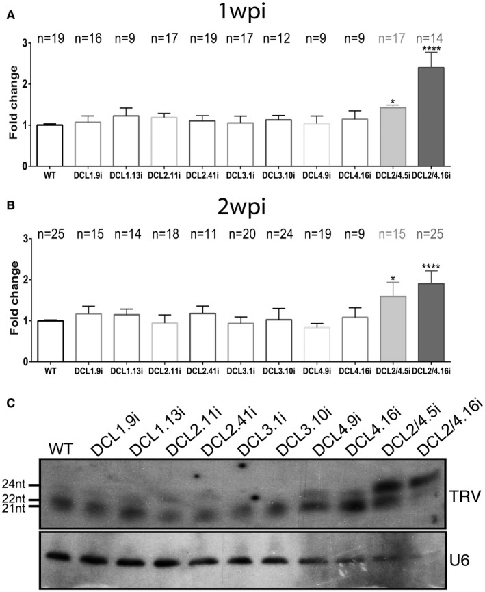
Figure 5.

Tobacco rattle virus (TRV)‐infected DCLi plants. Graphs representing measurements performed with Quantity One 4.4.1 (Biorad) in northern blots at 1 week post‐infection (wpi) (A) and 2 wpi (B). Statistical analysis was performed with one‐way analysis of variance (ANOVA) test, and the P value was set at *P < 0.05 and ****P < 0.0001. ‘n’ represents the number of individual measured plants. (C) Northern blot analysis of TRV vsiRNA from 2 wpi DCLi infected plants. U6 was used as loading control. WT, wild‐type.
