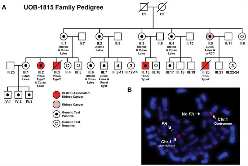
FIGURE 1.

Analysis of a large HLRCC family (UOB-1815) with a complete germline deletion of the FH gene. (A) A pedigree for a large HLRCC family that demonstrated several individuals with HLRCC associated type 2 papillary kidney cancer (patients highlighted in red). One individual (highlighted in pink) presented with clear cell kidney cancer that is currently not associated with HLRCC but is the most common sporadic kidney cancer. Additional features of HLRCC are listed under each individual if known. Solid black dots indicate complete loss of FH that has been confirmed by CLIA testing, while white dots with black outlines indicate testing confirmed no loss of FH . (B) An example of the FISH analysis of blood cells for a member of this pedigree that demonstrated germline loss of FH (green) near the q telomere of one copy of chromosome 1. Chromosome 1 was identified using a chromosome 1 centromere specific probe (red). [Color figure can be viewed at wileyonlinelibrary.com]
