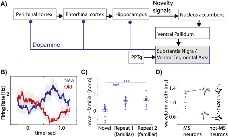
Figure 1: The Lisman Hippocampus VTA/SN loop model and novelty signaling human DA neurons.
(A) Schematic of interactions and flow of novelty signals within the hippocampus-VTA/SN loop. Adopted from [2, 3, 108]. (B-D) Novelty signaling human DA neurons. (B) Example neuron that increases its firing when a stimulus is novel (blue) and decreases when the same stimulus is shown again (red). (C) Population summary. Novelty-sensitive DA neurons change their firing rate between the first (left) and second (middle) time the same image is seen in a continuous recognition memory task. Each dot is one neuron. (D) Analysis of extracellular waveforms of neurons recorded in the human SN indicates a population of wide-and narrow waveform neurons, which are putatively dopaminergic and GABAergic, respectively. Note that the novelty-signaling neurons (blue) had wide waveforms. (B-D) adjusted from [24]. Abbreviations: SN – substantia nigra, VTA – ventral tegmental area, PPTg - pedunculopontine tegmental nucleus.

