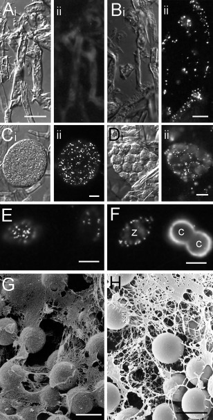
Figure 3.

Synthesis of secretory proteins during Phytophthora cinnamomi sporulation and their secretion during encystment. These processes are exemplified in (A)–(F) by immunolabelling with monoclonal antibody (mAb) PcCpa2 and in (G) and (H) by mucin‐like material visualized by scanning electron microscopy (SEM). Micrographs in (Ai)–(Di) are bright field images of the same field of view as shown in the fluorescence images in (Aii)–(Dii). (A) Cryosection of vegetative hyphae. No components in vegetative hyphae react with PcCpa2 mAb after immunofluorescence labelling. (B) Cryosection of sporulating hyphae immunolabelled with PcCpa2 mAb. PcCpa2 reacts with three high‐molecular‐weight polypeptides that are synthesized after the induction of sporulation and packaged into zoospore dorsal vesicles (Gubler and Hardham, 1988). (C) Mature sporangium immunolabelled with PcCpa2 mAb. Dorsal vesicles containing PcCpa2 proteins are randomly distributed throughout the sporangial cytoplasm. (D) During sporangial cleavage, the dorsal vesicles labelled by PcCpa2 mAb become distributed near cleavage membranes that will form the dorsal surface of the future zoospores. (E) PcCpa2‐containing vesicles next to the zoospore dorsal surface. (F) PcCpa2‐containing vesicles in the zoospore (z) cortical cytoplasm and on the surface of two young cysts (c). The absence of immunolabelling in the region of contact between the two cysts may be because this was the ventral surface of both cells or because the antibody did not have access to this region. (G) Mucin‐like material secreted during zoospore encystment on a root surface visualized by SEM after critical point drying. (H) Mucin‐like material secreted during zoospore encystment on a root surface visualized by cryo‐SEM. Images in (A)–(F) are courtesy of Dr Michele Cope. The image in (H) is reproduced with permission from Hardham et al. (1994). Bars, 10 µm.
