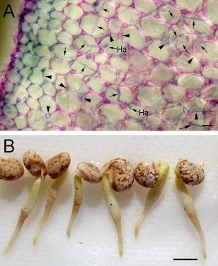
Figure 5.

Colonization and lesion development in lupin (Lupinus augustifolius) roots 20 h after inoculation with Phytophthora cinnamomi zoospores. (A) A transverse hand‐section of an infected lupin root. During initial colonization of the root, hyphae grow from the epidermis, through the cortex and into the vascular cylinder. Hyphal growth may be intracellular (arrows) or intercellular (arrowheads). Two putative haustoria (Ha) are indicated. (B) Lesions develop on the lupin roots just below the surface of the zoospore suspension whose approximate position is marked by black ink spots. Bars, 10 µm.
