Figure 7.

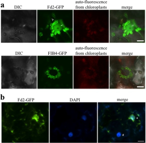
Fd2 localizes in stromules that extend from chloroplasts. (a) Confocal images of chloroplasts around the nuclei in Nicotiana benthamiana cells transiently expressing Fd2‐GFP (top) and FIB4‐GFP (bottom). Fd2, but not FIBRILLIN4 (FIB4), was found in stromules (top, indicated by arrowheads). A weak Fd2‐GFP signal was detected in the nucleus that was associated with the stromules (indicated by arrow). GFP, green fluorescent protein. (b) 4′,6‐Diamidino‐2‐phenylindole (DAPI) was used to visualize the nuclei. The nuclei that were connected with stromules showed strong Fd2‐GFP signals (indicated by *), whereas the nuclei without stromules attached showed very weak Fd2‐GFP signals (indicated by #). Bars, 10 μm (a) and 20 μm (b). DIC, Differential Interference Contrast.
