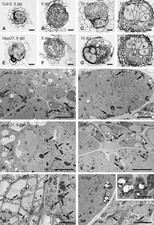
Figure 6.

Loss of function of HIPP27 causes physiological or metabolic abnormalities. Light (A–H) and transmission electron (I–O) microscopy images of cross‐sections of syncytia at 5 days post‐infection (dpi) (A, B, E, F, I, K and L) and 10 dpi (C, D, G, H, J and M–O) induced in roots of Col‐0 wild‐type plants (A–D, I and J) and hipp27a mutant (E–H and K–O). Light microscopy images were taken close to the nematode head (A, C, E and G) or through the widest regions of syncytia (B, D, F and H). Arrowheads indicate selected starch grains in plastids; arrows point to plastids. N, nematode; ND, neoplastic divisions; Pd, periderm; S, syncytium; SX, secondary xylem. Scale bars: 20 µm (A–H), 5 µm (I–O).
