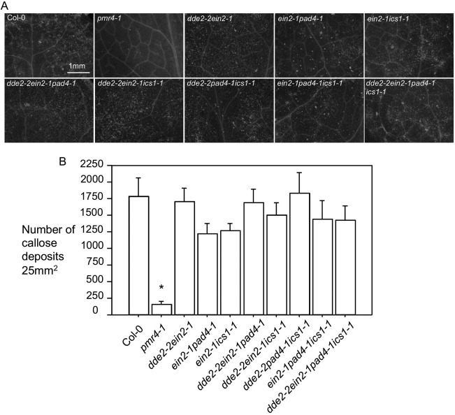
Figure 4.

Erwinia amylovora‐induced callose deposition in Arabidopsis is independent of salicylic acid (SA), ethylene (ET) and jasmonic acid (JA) signalling. Erwinia amylovora [optical density at 600 nm (OD600) = 0.01]‐infiltrated leaves of each genotype were harvested for fixation and staining with aniline blue. (A) Images of callose deposits. Callose deposits appeared as fluorescent dots and were photographed with an AxioCam MRc5 camera connected to a fluorescence stereoscope microscope. (B) Quantification of callose deposits. The number of callose deposits of each genotype was quantified using ImageJ (Version 1.45s). Each data point was an average of at least six images from four different leaves ± standard deviation. Statistical analysis was performed with one‐way analysis of variance (ANOVA) Fisher's partial least‐squares difference (PLSD) tests (StatView 5.0.1). The asterisk indicates significant difference of pmr4–1 from other genotypes (P < 0.05). These experiments were repeated twice with similar results.
