Figure 5.

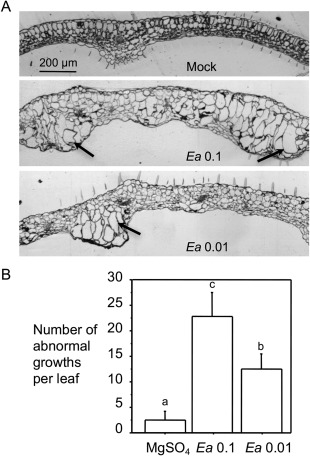
Erwinia amylovora induces abnormal cell growths in Arabidopsis. Erwinia amylovora‐infiltrated (Ea) and 10 mm MgSO4‐treated (Mock) leaves were observed for leaf morphology. (A) Leaf cross‐sections. The treated leaves were fixed, embedded in LR white resin and cut with an ultramicrotome into 1‐µm sections. Leaf cross‐sections were stained with 0.1% toluidine blue O and photographed using an AxioCam MRc5 camera (Zeiss, Inc., Göttingen, Germany) connected to a dissecting microscope. Arrows indicate enlarged cells in abnormal growth regions in leaves. The size bar represents 200 μm and applies to all images. (B) Quantification of the tumour‐like growths. The abnormal growths appeared to be transparent protrusions on the infected leaves and were counted at 5 days post‐inoculation (dpi) with the assistance of a dissecting microscope. At least 25 leaves were used for counting. Error bars represent standard deviation. Statistical analysis was performed with one‐way analysis of variance (ANOVA) Fisher's partial least‐squares difference (PLSD) tests (StatView 5.0.1). Different letters indicate significant difference amongst the samples (P < 0.05).
