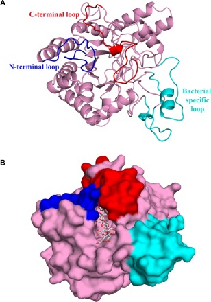
Figure 1.

Crystal structure of the catalytic domain of CbsA. (A) CbsA has a central distorted β‐barrel (pink) made up of eight strands. The N‐ and C‐terminal loops are shown in blue and red, respectively, whereas the unique loops present towards the C‐terminal region are shown in cyan. (B) The CbsA structure with the modelled substrate. The surface representation of the enzyme represents a well‐defined substrate‐binding tunnel responsible for processive hydrolysis of the single‐chain cellulose polymer. Three molecules of cellobiose were modelled from PDB ID: 4B4F.
