
Figure 4.
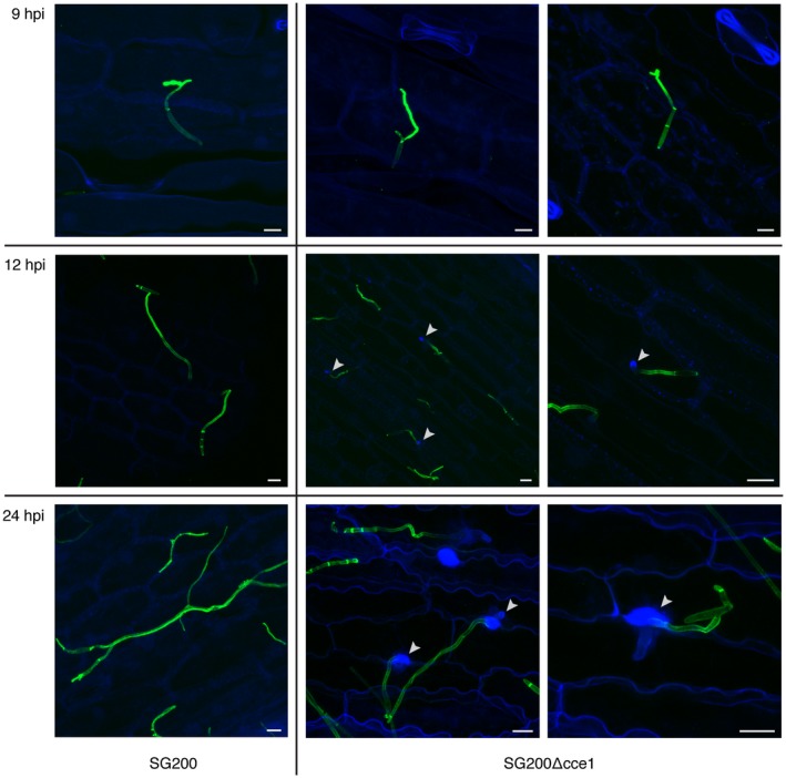
Detection of papillae formation by callose staining with aniline blue. The progenitor strain SG200 (left) does not induce callose deposition, whereas the SG200Δcce1 deletion strain (two right panels) causes papillae formation and callose deposition at penetration sites. Photomicrographs were taken 9, 12, and 24 hours post infection (hpi). WGA‐AF488 fluorescence (green) stains fungal hyphae; aniline blue fluorescence (blue) to visualize callose deposition; white arrows indicate sites of callose deposition. Scale bar = 10 μm.