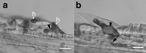
Figure 4.

Penetration of trichomes by Fusarium graminearum at 5 days post‐inoculation (dpi). (a) Infection of trichome companion cell from (b) with external hyphopodia (h) appressed to outside and thin penetration hypha (arrowhead). (b) Large prickle near paleal margin with fungal growth into hypodermis (arrow). Note: staining reveals silica crystal inside trichome (arrowhead). Bars, 20 μm.
