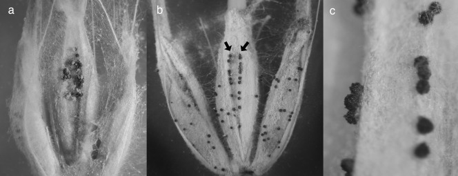
Figure 5.

Perithecia emerging from the surface of inoculated barley florets. (a) Perithecia at the location of the inoculation droplet on the palea of a floret infected before fusion. (b) Perithecia emerging from the paleal vascular bundle ridges (arrows), including beyond the location of infection (compare with Fig. 1a). (c) Detail of perithecia emerging from vascular bundle ridges.
