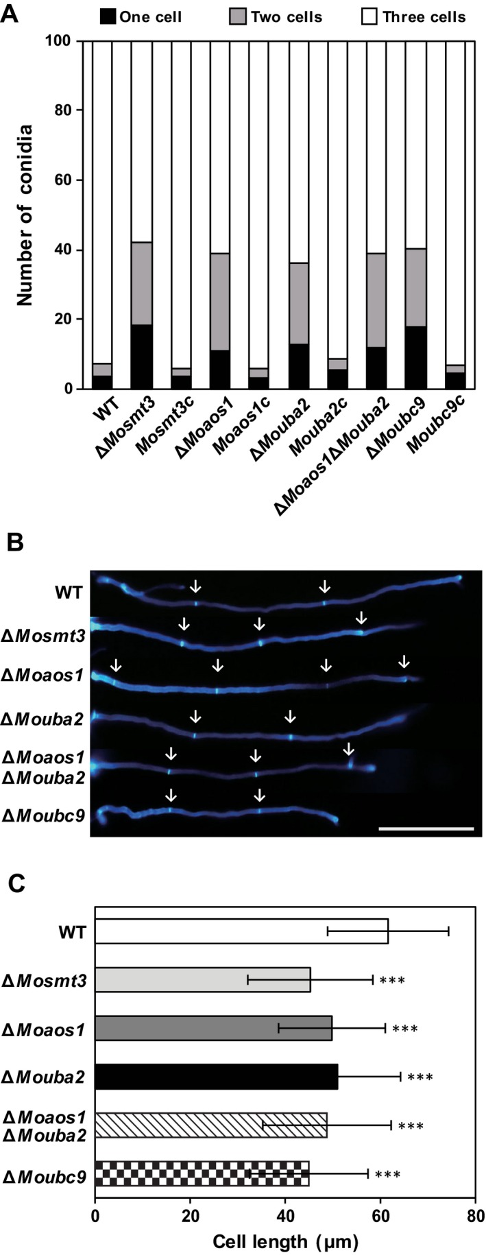
Figure 5.

Number of cells in conidia and cell length in hyphae. (A) Percentage of conidia with one, two or three cells in the wild‐type (WT), deletion mutants and complemented strains. (B) Hyphae were stained with Calcofluor white (CFW) and observed under a fluorescence microscope. White arrows indicate septa of hyphae; scale bar, 200 μm. (C) Cell length of hyphae was measured using ImageJ. Significance was determined by t‐test (***P < 0.001).
