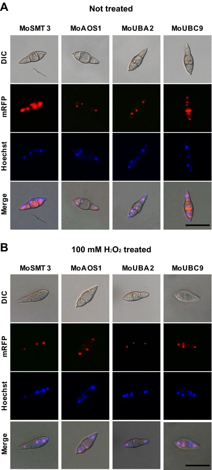
Figure 9.

Intracellular localization of SUMOylation components in Magnaporthe oryzae. (A) MoAOS1 and MoUBA2 were localized predominantly in the nucleus, but MoSMT3 and MoUBC9 were localized to both the nucleus and the cytoplasm. Nuclei were stained with Hoechst 33342. Scale bar, 25 μm. (B) The four SUMOylation components were predominantly localized to the nucleus under oxidative stress conditions (100 mm H2O2). Scale bar, 25 μm. Differential interference contrast (DIC), mRFP, monomeric red fluorescent protein.
