Figure 1.

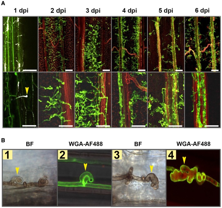
Ustilago hordei disease progression on barley leaves. (A) Wild‐type U. hordei (two mating types) was inoculated onto 10‐day‐old barley seedlings and disease progression was observed at 1, 2, 3, 4, 5 and 6 days post‐inoculation (dpi). Microscopic observations were performed with calcofluor white at 1 dpi and with WGA‐AF488/propidium iodide staining for the rest of the time points to visualize U. hordei colonization. Yellow arrow indicates U. hordei appressorium formation and host penetration. Scale bar at the top indicates 100 µm and at the bottom indicates 50 µm. Red represents plant cells and green represents fungal hyphae. (B) Ustilago hordei grows intracellularly in barley leaves and forms haustorium‐like structures during barley colonization. WGA‐AF488/propidium iodide staining and 3,3′‐diaminobenzidine (DAB) staining were performed to visualize U. hordei. Yellow arrows indicate clamps (photographs 1 and 2) and haustorium‐like structures (photographs 3 and 4). BF, bright field. The photographs were taken at 7 dpi. [Colour figure can be viewed at wileyonlinelibrary.com]
