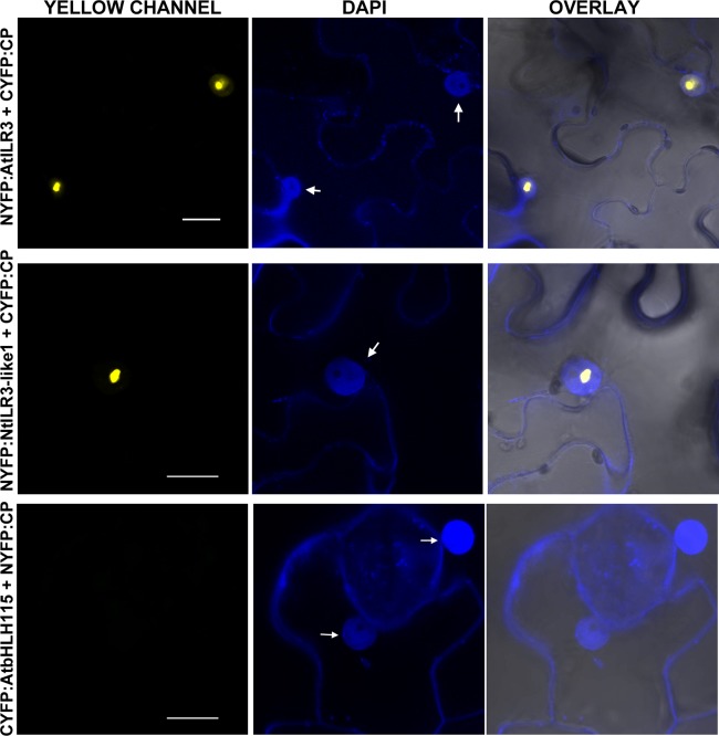
Figure 2.

Bimolecular fluorescence complementation (BiFC) analysis of coat protein (CP)–ILR3 interaction. Confocal laser‐scanning microscopy (CLSM) images of nuclei from epidermal cells co‐infiltrated with the constructs indicated on the left and stained with 4′,6‐diamidino‐2‐phenylindole (DAPI) are shown in the yellow (YELLOW, YFP) and blue (DAPI) channels. Overlay panels are the superposition of yellow fluorescent protein (YFP) and DAPI over the bright field images. YFP reconstitution was exclusively found in the nucleus (arrows). The BiFC nomenclature of the plasmids is as follows: NYFP and CYFP refer to the N‐terminal and C‐terminal fragments of YFP and, in all constructs, the YFP tag is attached to the N‐terminus of the protein. Bars, 10 µm.
