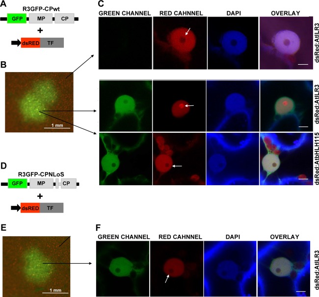
Figure 3.

Alfalfa mosaic virus (AMV) infection promotes nucleolar relocalization of AtILR3. (A, D) Schematic representation showing the modified AMV RNA3 with the wild‐type (wt) coat protein (CP) (R3GFP‐CPwt) or the mutated CP lacking the nucleolar localization signal (NLoS) (R3GFP‐CPNLoS) and expressing green fluorescent protein (GFP). (B, E) Images of one infection focus denoted by the accumulation of GFP. (C) Confocal laser‐scanning microscopy (CLSM) images in the green (GREEN), red (RED) and blue (4′,6‐diamidino‐2‐phenylindole, DAPI) channels of nuclei from non‐infected and infected cells with R3GFP‐CPwt and transiently expressing the transcription factors (TFs) indicated on the right of the panels. White arrows indicate the nucleolus. (F) CLSM images of a nucleus of a cell infected with R3GFP‐CPNLoS and transiently expressing dsRed:AtILR3. In this case, the TF does not accumulate in the nucleolus (white arrow). In all images, bar = 5 µm.
