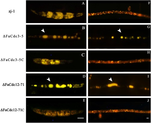
Figure 4.

Histochemical analysis of lipid droplets in conidia and hyphae. Lipid droplets in conidia (A–E) and hyphae (F–J) of the parental strain zj‐1, deletion mutants ΔFaCdc3‐5 and ΔFaCdc12‐71, and complemented strains ΔFaCdc3‐5C and ΔFaCdc12‐71C were stained with Nile red and examined using inverted fluorescence microscopy. Bar, 10 μm.
