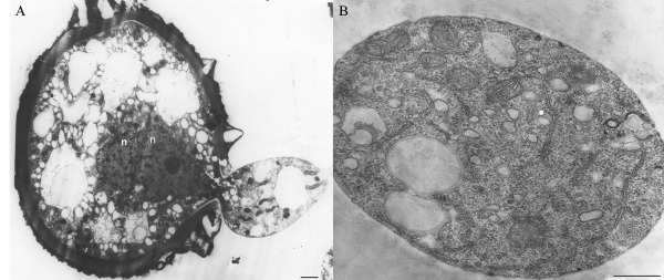
Figure 3.

Hemileia vastatrix urediniospore germination (Rijo and Sargent, 1983). (A) Beginning of germination, with cytoplasm passing through the germ pore with two nuclei (n) and one evident nucleolus (bar, 1 µm). (B) Transverse section of the germ tube showing different organelles (bar, 0.5 µm).
