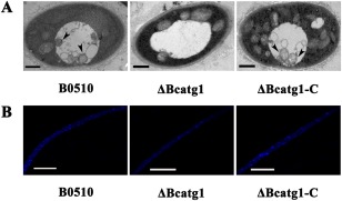
Figure 3.

Analysis of autophagy process by transmission electron microscopy (TEM) observation and monodansylcadaverine (MDC) staining. (A) After each strain had been incubated in nitrogen‐limiting medium [minimal medium without NaNO3 (MM–N)] with 2 mm phenylmethylsulfonylfluoride (PMSF) for 6 h, autophagosomes were evident in the vacuoles of the wild‐type strain B05.10 and △Bcatg1‐C, whereas no autophagosomes were observed in the vacuoles of △Bcatg1. Arrow, autophagosome. Scale bar, 0.5 μm. (B) Mycelia were stained with the fluorescent dye MDC and observed under epifluorescence microscopy; autophagy compartments were observed in B0510 and △Bcatg1‐C, but not in △Bcatg1. Scale bar, 10 μm.
