Figure 2.

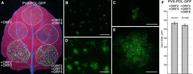
Complementation of cell‐to‐cell movement of transport‐deficient Potato virus X (PVX) derivative PVX‐POL‐GFP by Hibiscus green spot virus (HGSV) proteins in Nicotiana benthamiana leaves. (A) Green fluorescent protein (GFP) fluorescence in the leaf infiltrated with PVX‐POL‐GFP or PVX‐POL‐GFP in combination with the indicated HGSV protein‐encoding constructs. The leaf was imaged under UV light at 4 days post‐infiltration (dpi). Broken lines encircle infiltrated areas. (B–E) Fluorescence microscopy of infiltrated areas at low magnification (B, D) and higher magnification (C, E). (B, C) Images taken for PVX‐POL‐GFP. (D, E) Images taken for PVX‐POL‐GFP + ORF2 + ORF3. Scale bars: (B, D) 500 μm; (C, E) 200 μm. (F) The mean sizes of fluorescent foci observed in leaf areas in which PVX‐POL‐GFP was co‐agroinfiltrated with either ORF2 + ORF3 or ORF2 + ORF3 + ORF4 construct. ORF, open reading frame.
