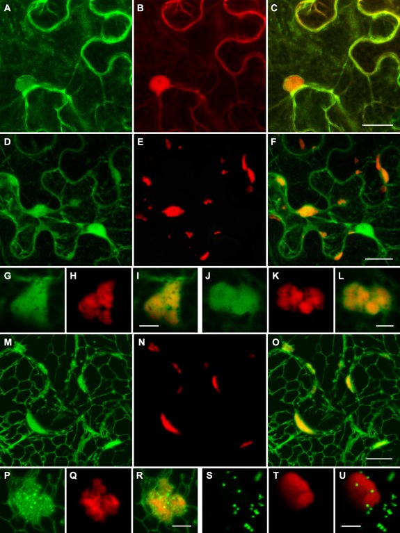
Figure 4.

Subcellular localization of GFP‐BMB1 and BMB2‐mRFP. (A–C) Co‐expression of GFP‐BMB1 and mRFP. (D–L) Co‐expression of BMB2‐mRFP and GFP. (M–R) Co‐expression of BMB2‐mRFP and ER‐GFP. (S–U) Co‐expression of BMB2‐mRFP and the Golgi marker ST‐GFP. Higher magnification images in (G–L) show the localization of the BMB2‐mRFP bodies in GFP‐containing cytoplasmic areas, and in (P–R) show fine granular ER‐GFP‐containing structures associated with the BMB2‐mRFP bodies. GFP and mRFP fluorescence signals were imaged independently. Merged images are shown in (C), (F), (I), (L), (O), (R) and (U). All images are reconstructed from Z‐series of optical sections. Scale bars: (C, F) 20 μm; (O) 10 μm; (I, L, R, U) 5 μm. BMB, binary movement block; ER, endoplasmic reticulum; GFP, green fluorescent protein; mRFP, monomeric red fluorescent protein.
