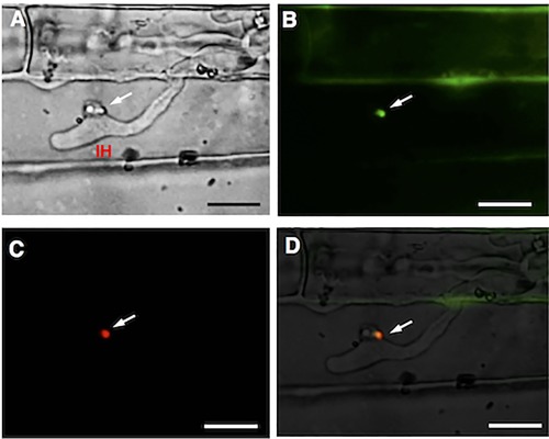
Figure 7.

Co‐localization of AVR‐Pia and PWL2 in the biotrophic interfacial complex (BIC) structures of Magnaporthe oryzae. Microscopic observation of rice sheath cells infected with Ina168m95‐1‐pCSN43‐DEST‐PPR::AVR‐Pia::4GGS::eGFP‐pPWL2::PWL2::mRFP‐transformed M. oryzae was performed at 57 h post‐inoculation (hpi). IH, invasive hypha; white arrows, BICs. Scale bars, 10 μm. (A) Bright‐field image. (B) Green enhanced green fluorescence protein (eGFP)‐labelled AVR‐Pia fluorescence in BICs. (C) Red monomeric red fluorescent protein (mRFP)‐labelled PWL2 fluorescence in BIC. (D) Merged bright‐field, eGFP::AVR‐Pia (green) and mRFP::PWL2 (red) images.
