
Figure 2.
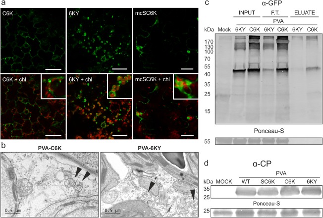
PVA‐C6K and PVA‐6KY are both infectious and the N‐terminal 6K2 tag is accessible for affinity purification. (a) Comparison of fluorescence derived from C6K (green) and 6KY (green) during Potato virus A (PVA) infection by confocal microscopy. The C6K protein localized mostly to scattered vesicles, whereas the 6KY signal was detected mostly in association with chloroplasts (chl., red). Magnified sections show 6K2 vesicle association with chloroplasts. (b) Electron microscopic images of the infected tissues. Both PVA‐C6K and PVA‐6KY produced cytoplasmic cylindrical inclusions which are indicated with arrowheads. (c) Affinity chromatography purification of the 6K2‐fusion protein from the infection context, revealing N‐terminally fused Cerulean fluorescent protein (CFP) to be better accessible for the green fluorescent protein (GFP)‐trap matrix compared with the C‐terminally fused yellow fluorescent protein (YFP). (d) Western blot analysis verified the presence of PVA coat protein (CP) in the upper leaves at 10 days post‐infiltration (DPI), indicating that PVA‐SC6K, PVA‐C6K and PVA‐6KY are all able to cause systemic infection.