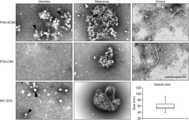
Figure 5.

Morphological characterization of the purified 6K2‐associated membranes. Affinity‐purified PVA‐SC6K, PVA‐C6K and MC‐SC6K samples were negatively stained with uranyl acetate and examined by electron microscopy (EM). Two types of membrane structure were observed: individual vesicles (left panels; shown by arrowheads) and membrane clusters (middle panels). Strep‐Tactin matrix captured PVA particles non‐specifically from infected samples (right panels). The sizes of the purified individual vesicles vary between 50 and 100 nm, with the median size being 56 nm (n = 40). Scale bar, 500 nm.
