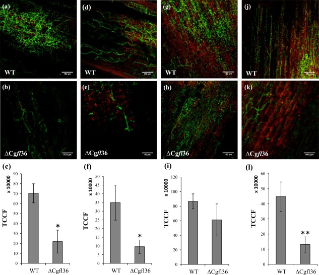
Figure 5.

Live cell confocal imaging of maize leaves infected with wild‐type (WT) Colletotrichum graminicola and the ΔCgfl transformants. (a) Fungal infection in maize leaves was monitored by confocal microscopy at 60 h post‐infection (hpi) and 96 hpi. A detailed examination of the lesions at 60 hpi showed that the infection proceeded more slowly in the ΔCgfl36 strain relative to the WT strain, indicating that the fungus had reduced ability to colonize host maize cells in the central zone (a, b) and in the borders of the lesion (d, e) with respect to the WT strain. Differences in fungal growth were quantified by measuring green fluorescent protein (GFP) fluorescence (c, f) using the software ImageJ. The values shown in the graph represent averages of total corrected cellular fluorescence (TCCF) of two independent experiments with standard deviation (SD) bars. Asterisk denotes samples that were significantly different from the WT (P < 0.05). At 96 hpi, both WT and ΔCgfl36 strains showed similar growth in the central zone of lesions (g, h) but, in the lesion borders, the ΔCgfl36 mutant exhibited delayed development (j, k). Fungal growth was quantified by measuring GFP fluorescence, and TCCF values are represented in graphs (i) and (l). Double asterisks denote samples that were significantly different from the WT (P < 0.001).
