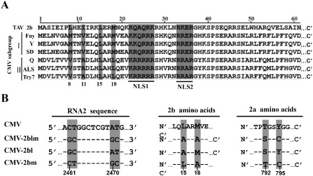
Figure 1.

Alignment of 2b protein sequences and schematics showing Cucumber mosaic virus (CMV) 2b point mutants. (A) Alignment of 2b protein sequences from Tomato aspermy virus (TAV) and different CMV subgroup I and II isolates. All of the sequence alignments are from the N‐terminal 61 amino acids of the diverse 2b derivatives. The two nuclear localization signal (NLS) sequences are highlighted in grey and underlined. The N‐terminal eighth, 11th, 15th and 18th amino acids are indicated in grey. (B) The nucleotide substitutions (left panel) in the CMV‐2blm, CMV‐2bl and CMV‐2bm virus mutants and the resulting amino acid point mutations in the 2b (middle panel) and 2a (right panel) regions. The point mutations are highlighted with a grey background and their positions in the genome sequences are indicated at the bottom.
