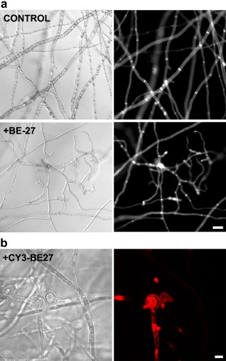
Figure 2.

(a) Morphological changes in Penicillium digitatum mycelium exposed to BE27. Penicillium digitatum mycelium was grown in the absence (control) or presence of 5 μg/mL of BE27. After 24 h of incubation, samples were stained with calcofluor white (CFW) and visualized under the microscope. Panels represent bright‐field images (left) and fluorescence images indicative of CFW staining (right) for the same fields. Bar, 20 μm. (b) Interaction between BE27 and P. digitatum. To visualize the interaction of BE27 with fungal structures, conidia of P. digitatum were grown in vitro in potato dextrose broth (PDB) medium for 24 h before exposure to 5 μg/mL of CY3‐BE27 for 24 h. After washing, the samples were visualized with a confocal laser microscope. The red channel image (right panel) and the corresponding interference contrast image (left panel) are shown. Bar, 5 μm.
