Figure 5.

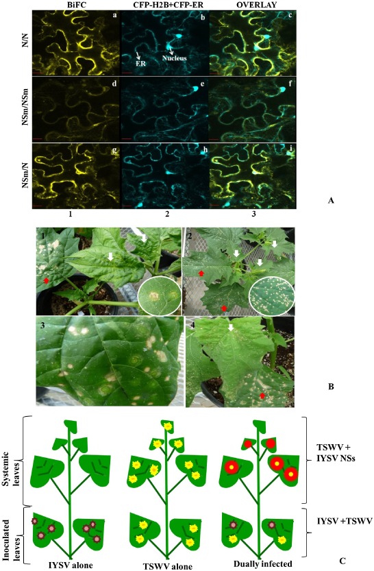
(A) Confocal micrographs showing the Iris yellow spot virus (IYSV) protein interactions examined. Interaction assays were performed in leaf epidermal cells of transgenic Nicotiana benthamiana plants expressing cyan fluorescent protein fused to the nuclear marker histone 2B (CFP‐H2B) and the endoplasmic reticulum marker (CFP‐ER). Shown are the interaction assay [bimolecular fluorescence complementation (BiFC), column 1], localization of CFP‐H2B and CFP‐ER (nucleus and ER, column 2) and a merge of all panels (overlay, column 3). Proteins listed first in the pair of interactors were expressed as C‐terminal fusions to the amino‐terminal half of yellow fluorescent protein (YFP). Those listed second were expressed as C‐terminal fusions to the carboxy‐terminal half of YFP. All protein fusions to each half of YFP were tested in all pairwise combinations, a subset of detectable interactions of which is shown here: (a–c) N/N; (d–f) NSm/NSm; (g–i) NSm/N. Micrographs shown are representative of at least 50 cells examined. Scale bar, 20 μm. (B) Differential response of Datura stramonium to mechanical inoculation with IYSV and Tomato spotted wilt virus (TSWV). Top left: D. stramonium is a permissive host for TSWV, wherein infection results in concentric ring spots in inoculated leaves, and leaf curling, mottling and necrosis in systemic leaves, shown by white arrows. Top right: D. stramonium is a restrictive host for IYSV. IYSV infection produces small necrotic spots. Bottom left: distinctive concentric rings and necrotic spots develop in inoculated leaves of Datura infected with both viruses. Bottom right: more severe symptoms develop in inoculated and systemic leaves of dually infected Datura. Red arrows show symptoms in inoculated leaves and white arrows show symptoms in systemic infection. Insets show close‐up of TSWV and IYSV symptoms in inoculated leaves. (C) Schematic representation of differential response of D. stramonium to IYSV and TSWV.
