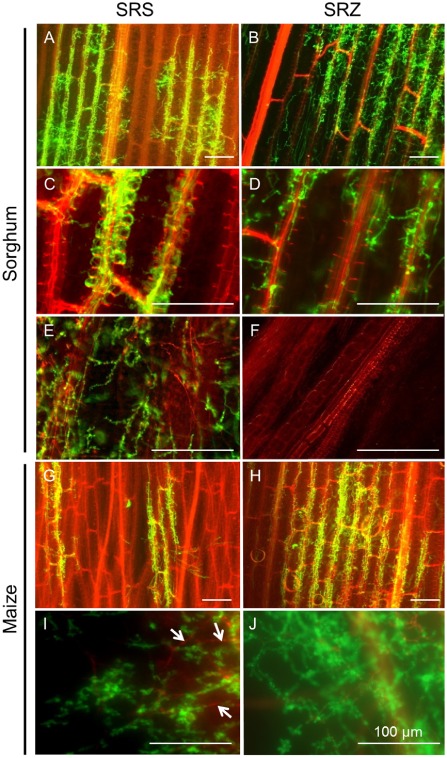
Figure 1.

Microscopic characterization of sorghum and maize infection by S porisorium reilianum. Sorghum (A–F) and maize (G–J) seedlings were syringe inoculated with S . reilianum f. sp. reilianum (SRS) (left) or S . reilianum f. sp. zeae (SRZ) (right). Samples were collected at 4 days after inoculation (dai) (A, B, G, H), 9 dai (C, D) or 15 dai (E, F, I, J). Plant material and dead hyphae were stained with propidium iodide and appear red; fungal hyphae were stained with wheat germ agglutinin (WGA)‐Alexafluor‐488 and appear green. Sorghum samples inoculated with SRS show hyphae colonizing leaf tissues (A) and presenting a preference for vascular bundles (C), later reaching the nodes and apical meristems (E). SRZ infects leaves (B) and leaf sheaths (D), without showing such a preference for vascular bundles, and without reaching the nodes and apical meristems (F). Maize plants inoculated with SRS show hyphae colonizing leaf tissues (G) and reaching the nodes and apical meristem (I), where some dead hyphae are also observed (white arrows). Leaves inoculated with SRZ show extensive fungal growth (H) and the pathogen reaches the nodes and apical meristem (J). Bars, 100 μm; n = 12 plant samples.
