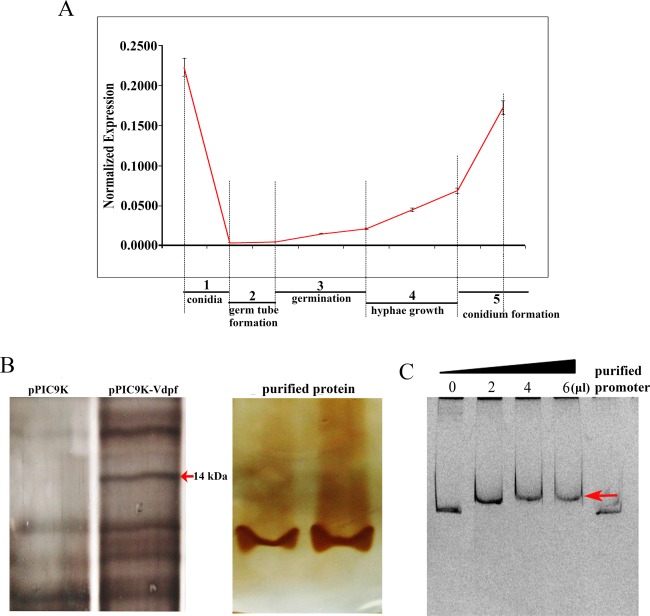
Figure 6.

Normalized expression of Vdpf at different stages and in the electrophoretic mobility shift assay (EMSA) of Vdpf. (A) The relative transcript amount at each stage was determined in comparison with the 18srRNA transcripts in the same tissue. The y axes illustrate the relative quantity of the transcripts compared with the housekeeping gene: 18srRNA. Three biological replicates (n = 3) were used for this study. The error bars indicate the standard deviation. The statistical analysis was performed using the t‐test. 0 h, conidia; 6–12 h, formation of germ tube; 12–24 h, conidia germination; 24–48 h, hyphal growth; 48–120 h, conidium formation. (B) The Zn(II)2Cys6 domain expressed in Pichia pastoris (Gs115). The protein expressed in Pichia pastoris was transformed with purified pPIC9k and served as the control. Vdpf (EGY18187.1) was purified using His‐tag. (C) Vdpf binds to its own promoter. A 1000‐bp promoter fragment of Vdpf was used for binding assays with 0, 2, 4 or 6 μL of purified Vdpf. The concentration of purified Vdpf was 513.636 μg/mL. The pure promoter was used as a probe control. The red arrow points to the shift bands. Three biological replicates (n = 3) were used for this study.
