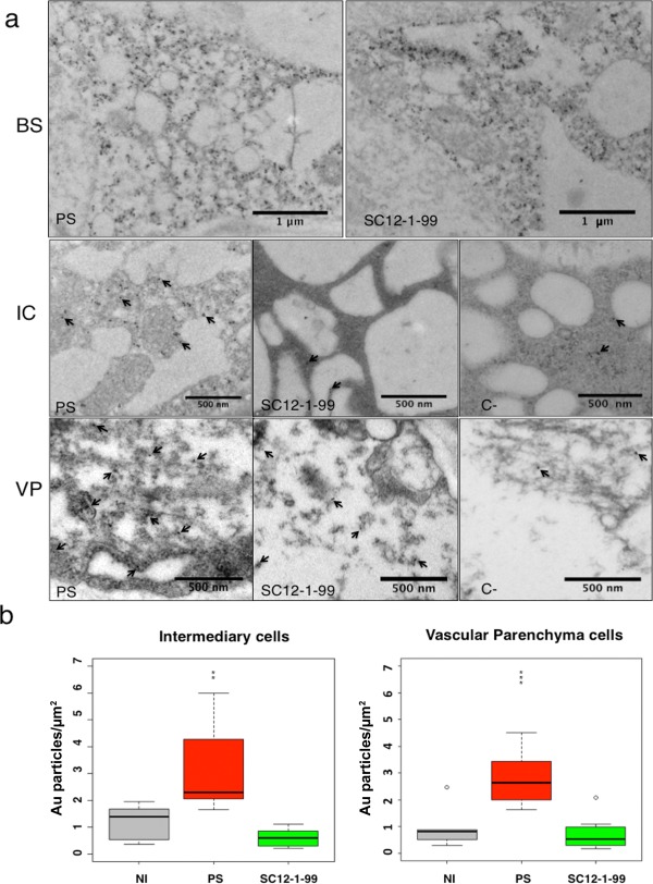
Figure 4.

Localization of CMV‐LS coat protein (CP) in the cells of minor veins. (a) Transmission electron microscopic immunocytochemistry of Piel de Sapo (PS) and SC12‐1‐99 lines inoculated with CMV‐LS. Gold particles are detected in bundle sheath (BS) cells, intermediary cells (ICs) or vascular parenchyma (VP) cells. In ICs and VP cells, arrows point to some gold particles (10 nm) in the sections. C‐, negative control from a non‐inoculated PS plant. PS and SC12‐1‐99 are susceptible and resistant lines, respectively. (b) Quantification of gold particles in ICs and VP cells of PS and SC12‐1‐99 lines. Three grids per sample type were used for counting. The numbers of gold particles in ICs and VP cells are represented as Au particles/μm2. The data were analysed using Student's t‐test and were considered to be significant when P ≤ 0.05. Results are represented in a box plot. The bottom and top of the box are the first and third quartiles, respectively. The band inside the box is the median, the second quartile. Outliers or individual points represent the variability outside the upper and lower quartiles. NI, non‐inoculated plants indicating non‐specific gold labelling. Significant differences between PS and SC12‐1‐99 are represented by asterisks: **P ≤ 0.01; ***P ≤ 0.001. CMV, Cucumber mosaic virus.
