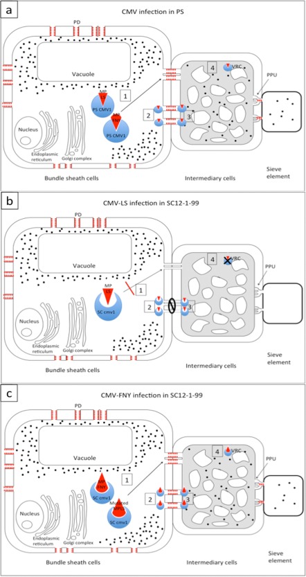
Figure 6.

Hypothetical model of CMV‐LS and CMV‐FNY infecting Piel de Sapo (PS) and SC12‐1‐99. The movement protein (MP) (red triangles) opens the size exclusion limit of plasmodesmata (PDs) surrounding the bundle sheath (BS) cells. In all cases, virus (black dots) accumulates in the BS cells. For simplicity, only traffic between BS cells and intermediary cells (ICs) is represented. The model would be the same for traffic between BS cells and vascular parenchyma (VP) cells. (a) Cucumber mosaic virus (CMV) infecting PS. In the BS cells, the protein PS CMV1 can interact directly or indirectly with MPs of both CMV‐LS and CMV‐FNY, leading either to transport to PDs (1) or to the opening of PDs (2), allowing systemic spread of the virus. In (3), the interaction between MP and CMV1 in the ICs can allow viral RNA to enter the ICs. Alternatively, the interaction MP–CMV1 could take place in the viral replication complexes (VRCs) of the ICs, allowing viral replication (4). (b) SC12‐1‐99 infected with CMV‐LS. LS MP would not be able to interact with SC cmv1 protein, impeding all of the above four possibilities. (c) CMV‐FNY or mutated CMV‐LS infection of SC12‐1‐99. FNY MP and mutated LS MP carrying the relevant residues (Guiu‐Aragonés et al., 2015) are able to interact with the SC cmv1 protein and allow any of the four possibilities given above. PPU, plasmodesmata pore units.
