
Figure 6.
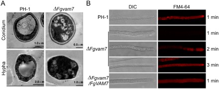
The ΔFgvam7 mutant was defective in vacuole morphogenesis and endocytosis. (A) The vacuole structure of the ΔFgvam7 mutant conidium and young hypha shown by transmission electron microscopy. (B) FM4‐64 staining assay. Strains were grown for 1 day on PDA (46 g potato dextrose agar powder in 1 L double‐distilled H2O)‐overlaid microscope slides before the addition of FM4‐64. Photographs were taken at the indicated periods. DIC, differential interference contrast.