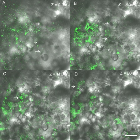
Figure 3.

Colonization of bacterial spot Xanthomonas perforans in the tomato phyllosphere. Bacterium enters though stomata (A), followed by growth in the substomatal chamber (B) (indicated by arrow). Bacterium multiplies in the intercellular spaces (C and D). Representative photomicrographs showing green fluorescent protein (GFP)‐labelled virulent X. perforans aggregates that are part of the lesion along a Z stack of 20 μm overlaid with Nomarski differential interference contrast images (40× magnification). Z values represent the distance in micrometres from the abaxial (lower) leaf surface. White bar, 25 μm.
