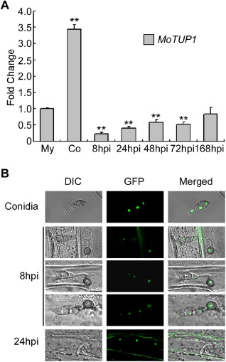
Figure 2.

Phase specific expression and subcellular localization of MoTUP1. A. The phase specific expression of MoTUP1 was quantified by quantitative real‐time PCR with synthesis of cDNA from each sample including infectious growth, vegetative growth and conidia. Relative abundance of MoTUP1 transcripts during infectious growth (from ungerminated conidia to in planta fungal cells 168 hpi) was normalized by comparing with vegetative growth in liquid CM (Relative transcript level = 1). Three independent biological experiments with three replicates in each were performed. The double asterisks represent significant difference between other different stages and mycelia stage (p < 0.01). My: Mycelia; Co: Conidia; hpi: hour post inoculation. B. MoTup1 is localized to the nucleus of the conidia in the complemented strain. The subcellular localization of MoTup1 during appressorium and invasive growth was performed by placing conidial suspensions on barley epidermis, and photographs were taken at 8 h and 24 h after incubation, respectively.
