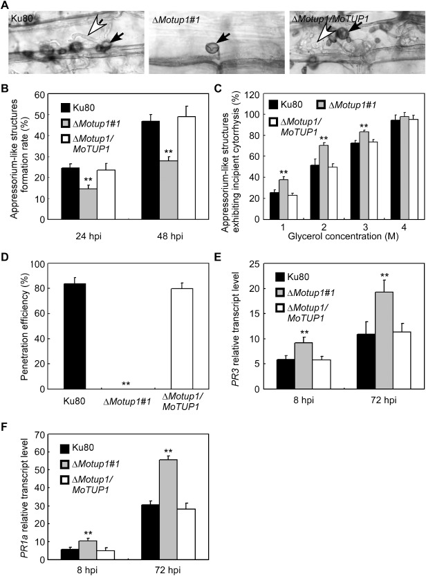
Figure 8.

Rice epidermis cell penetration assay. A. The mycelia of Ku80, ΔMotup1 mutant and the complemented strain were inoculated on the rice leaf tissue for 48 h. The rice epidermis cell penetration was observed under light microscope. White arrows point to invasive hyphae; black arrows point to appressorium‐like structures. B. The mycelia of Ku80, ΔMotup1 mutant and the complemented strain were inoculated on the barley leaf epidermis for 24 and 48 h, respectively. The formation rate of appressorium‐like structures was measured under light microscope. C. Quantification of collapsed appressorium‐like structures of Ku80 and the ΔMotup1 mutant strains. For each glycerol concentration, at least 50 appressorium‐like structures were observed and numbers of collapsed appressorium‐like structures were counted. D. Percentage of appressorium‐like structures penetrated and formed invasive hyphae on barley leaves 48 hpi. E and F. The transcription of PR3 and PR1a in the infected host was assayed using qRT‐PCR. RNA samples were collected from rice plants (Oryza sativa cv. CO‐39) 0, 8 and 72 h after inoculation with the ΔMotup1 mutant or the wild‐type strain. The average threshold cycle of triplicate reactions was normalized by the stable‐expressions gene elongation factor 1a (Os03g08020) in O. sativa. Three independent biological experiments with three replicates each time were performed and similar results were obtained in each time. Double asterisks represent significant differences (p < 0.01).
