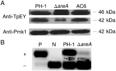
Figure 7.

Mitogen‐activated protein kinase (MAPK) phosphorylation and protein kinase A (PKA) activity assays. (A) Assays for the activation of Mgv1 and Gpmk1 MAPKs. Total proteins were isolated from vegetative hyphae of the wild‐type PH‐1, ΔareA mutant and complementation strain (AC6). The phosphorylation of Mgv1 (46 kDa) and Gpmk1 (42 kDa) was detected with the anti‐TpEY antibody. The expression level of Gpmk1 was detected with the anti‐Pmk1 antibody. (B) PKA activities were assayed with proteins isolated from hyphae of PH‐1 and ΔareA mutant using the PepTag A1 PKA substrate peptide. Whereas phosphorylated peptides migrated towards the anode (+), unphosphorylated peptides migrated towards the cathode (–) on a 0.8% agarose gel. N, non‐phosphorylated sample control; P, phosphorylated sample control.
