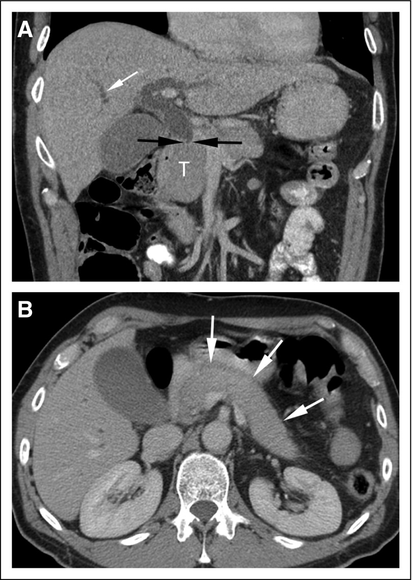
FIG 1.
Coronal (A) and axial (B) contrast-enhanced computed tomography (CT) images. (A) The coronal image demonstrates abrupt cutoff of the common bile duct (black arrows) at the level of the enlarged pancreatic head (T), with intrahepatic biliary duct dilatation (white arrow). The study was initially interpreted as demonstrating a 3.9-cm cancer in the pancreatic head. The patient was scheduled for a Whipple procedure but decided to seek a second opinion before surgery. At the request of the consulting surgeon, the same set of CT images was reviewed by a specialized radiologist who participates in a hepato-pancreato-biliary tumor board. Although the head of the pancreas is enlarged, the axial CT image (B) also shows diffuse pancreatic enlargement (arrows), with no visible pancreatic duct. The imaging findings were thus considered suspicious for autoimmune pancreatitis, and measurement of circulating IgG4 levels was recommended. Subsequent testing revealed elevated IgG4 levels. Response to steroid therapy confirmed the diagnosis of autoimmune pancreatitis. Images courtesy of Richard Do, MD, PhD, Memorial Sloan Kettering Cancer Center.

