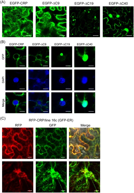
Figure 5.

Subcellular distribution of wild‐type and deletion mutants of Chinese wheat mosaic virus (CWMV) cysteine‐rich protein (CRP). (A, B) Epidermal cells of Nicotiana benthamiana plants transiently expressing enhanced green fluorescent protein (EGFP) fused with CWMV CRP wild‐type or mutants. Panels in (B) show the perinuclear areas. (C) Subcellular distribution of red fluorescent protein (RFP) fused with CWMV CRP in epidermal cells of transgenic N. benthamiana line 16c expressing GFP. Top and bottom panels show the cytoplasmic and perinuclear areas, respectively. Fluorescent proteins and 4′,6‐diamidino‐2‐phenylindole (DAPI) staining were observed using confocal laser scanning microscopy (CLSM) at 3 days after inoculation (dai). Images are derived from single confocal sections. Bars: 20 μm (A); 10 μm (B, C).
