Figure 2.

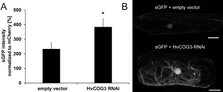
Analysis of green fluorescence protein (GFP) secretion in cells expressing the HvCOG3 RNA interference (RNAi) construct. GFP was N‐terminally fused with a signal peptide. The resulting construct (sGFP in pGY‐1) was transiently co‐transformed, together with either the empty vector pIPKTA30N as a control or the HvCOG3 RNAi construct and the transformation marker mCherry, into epidermal cells of detached barley leaves (cv. ‘Golden Promise’) and analysed by confocal laser scanning microscopy 2 days after transformation. (A) The intensity of sGFP in the cytoplasm of transformed cells was normalized against the fluorescence intensity of mCherry. In each of three independent repetitions, 50 cells were examined. All cells were imaged with the same excitation and detection settings. To warrant detection within the dynamic range of the system, the brightly fluorescing mCherry was recorded at low detector gain, whereas weakly fluorescing sGFP was recorded at higher detector gain. Average GFP fluorescence in HvCOG3 deficient cells is significantly higher than in control cells (Student's t‐test, P < 0.05). (B) Confocal laser scanning micrographs of barley epidermal cells expressing sGFP together with either the empty vector (top) or the HvCOG3 RNAi construct (bottom) 2 days after transformation. Photographs were taken using the same detection settings. Size bars, 20 μm.
