
Figure 4.
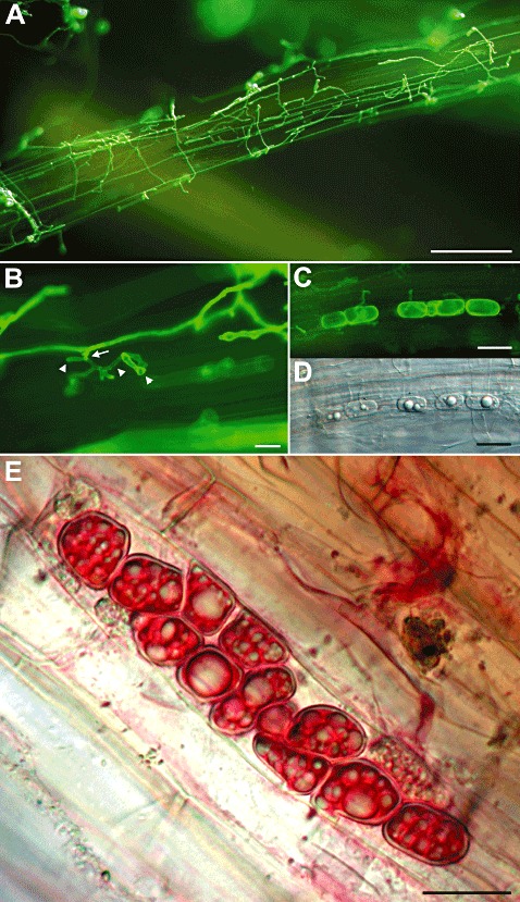
Colonization of Arabidopsis root by Piriformospora indica. (A) Large areas of the root's maturation zone are colonized by P. indica at 7 days after inoculation (dai). (B) Root colonization is achieved by direct cell penetration (arrow) and by the subsequent establishment of intracellular hyphae (arrowheads). (C, D) Root colonization is associated with the development of intracellular spores in colonized cells. (C) Fluorescence image of intracellular spores. (D) Bright field image of (C). (E) Chlamydospores of P. indica in tomato root cells. Fungal structures were stained with WGA‐AF488 (Invitrogen, Darmstadt, Germany) and visualized by fluorescence microscopy (excitation/emission: 470–520/505–530 nm) using an Axioplan 2 Microscope (Zeiss, Jena, Germany) (A–C), or stained with fuchsin–lactic acid and visualized by bright field microscopy (E). Bars: 60 µm (A); 10 µm (B–E).