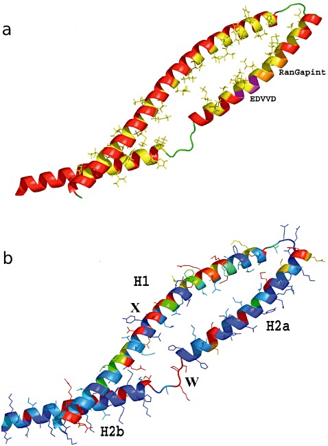
Figure 7.

LR10 coiled coil (CC) model. (a) Diagram coloured by secondary structure; amino acids involved in hydrophobic zipping are coloured yellow to pinpoint the hydrophobic matching between helices. The RanGAP interacting motif and the EDVVD motif are coloured in orange and magenta, respectively (Rairdan et al., 2008; Slootweg et al., 2010; Tameling and Baulcombe, 2007). (b) Diagram with variability mapped on a scale from blue (conserved) to red (hypervariable); X, the exposed surface of the first strand (H1); W, the linkers between H2a and H2b.
