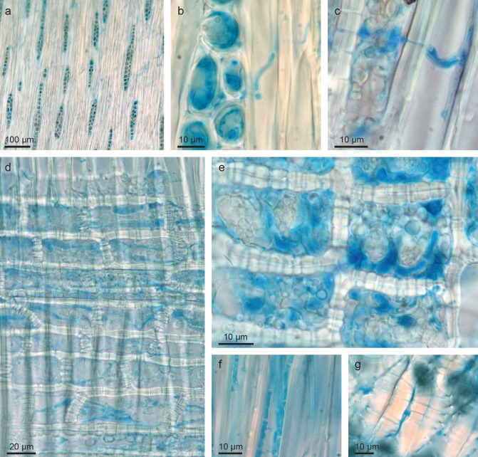
Figure 3.

Mycelium of Hymenoscyphus pseudoalbidus in Fraxinus excelsior tissues (stained with lactophenol blue): (a, b) hyphae concentrated in xylem rays (tangential section); (c) hypha growing through a cell wall separating a ray cell from a fibre cell; (d, e) hyphae in xylem ray cells (radial section); (f) hyphae growing in a xylem fibre in an axial direction; (g) hyphae growing within the lumen and pit channels of a bark sclereid.
